Medical Imaging Without Radiation
Context
Scoliosis, or deformity of the spinal column, affects 2% to 4% of the population. It is detected early: starting in adolescence, patients are checked regularly so the curvature of the spine can be observed as it develops. On average, young patients are x-rayed every 3 to 6 months, which exposes them to radiation. Even though the HUG is equipped with a low radiation x-ray system (EOS System), these repeated exposures to radiation can be harmful for a young patient, given their regular frequency.
Project
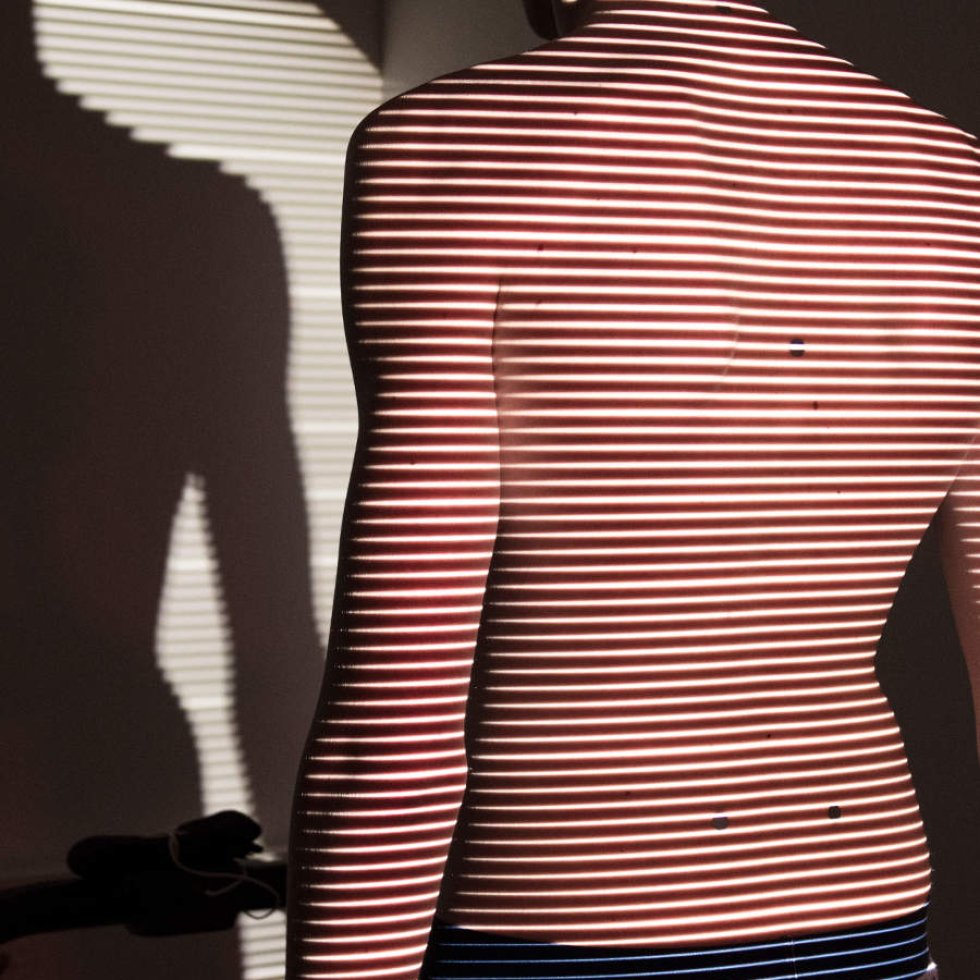
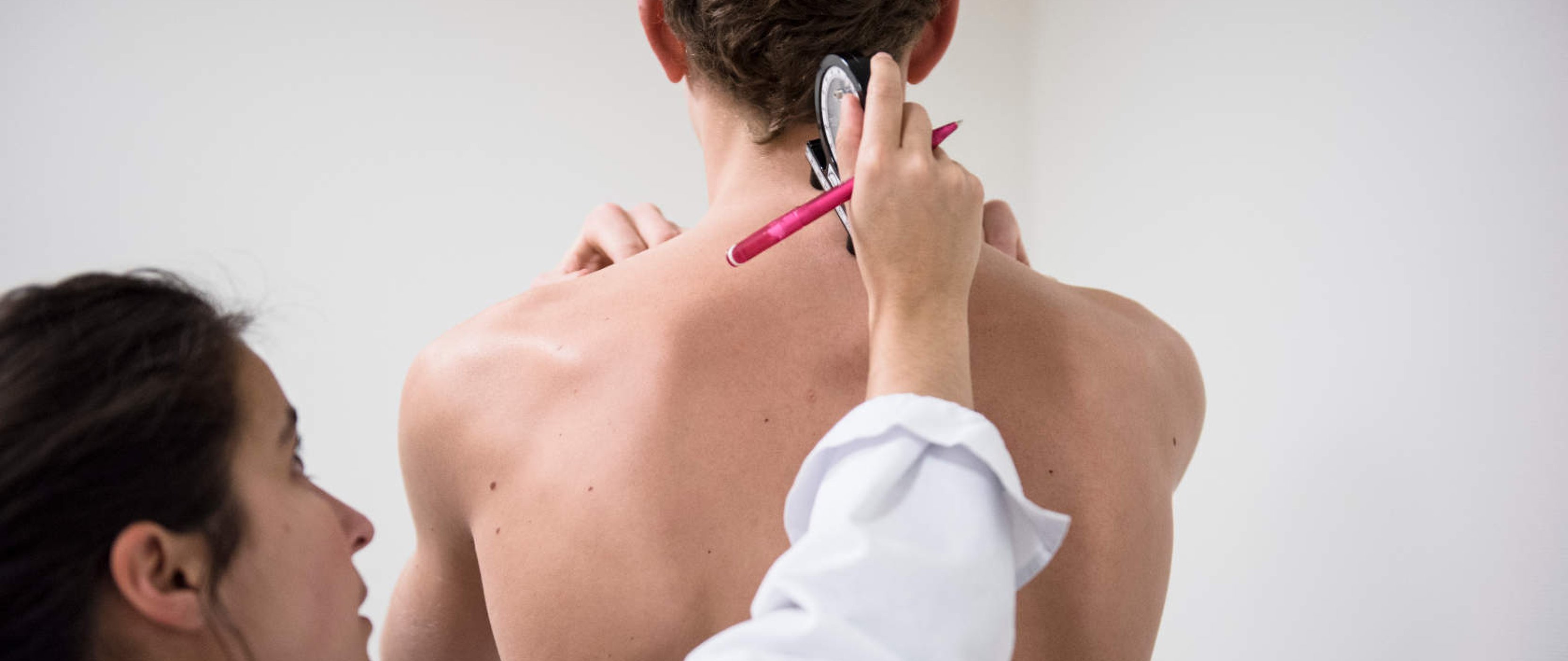
The project is for use of a 4D photography device, called Raster, to monitor young patients with scoliosis. This system, already in use at the HUG, does not use radiation and enables 4D imaging of the spinal column, the 4th dimension being patient movement. We will implement a systematic procedure of evaluating these young patients using the 4D Raster system in comparison with the traditional EOS x-ray system. If the parameters obtained by the two systems are well-correlated, this new practice will be systematically implemented.
project manager
Dr. Romain Dayer, Deputy Head Physician and Clinical Professor, Pediatric Orthopedics Division, Children and Adolescents Department, Geneva University Hospitals
Galerie photo